Artificial Organs May Finally Get a Blood Supply
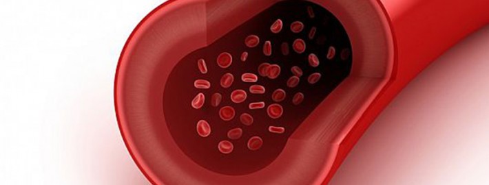
Artificial tissue has always lacked a key ingredient: blood vessels. A new 3-D printing technique seems poised to change that.
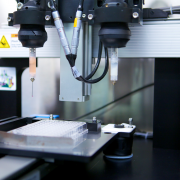
Using a custom-built four-head 3-D printer and a “disappearing” ink, materials scientist Jennifer Lewisand her team created a patch of tissue containing skin cells and biological structural material interwoven with blood-vessel-like structures.Reported by the team in Advanced Materials, the tissue is the first made through 3-D printing to include potentially functional blood vessels embedded among multiple, patterned cell types.
In recent years, researchers have made impressive progress in building tissues and organ-like structures in the lab. Thin artificial tissues, such as a trachea grown from a patient’s own cells, are already being used to treat patients (see “Manufacturing Organs”). In other more preliminary examples, scientists have shown that specific culture conditions can push stem cells to grow into self-organized structures resembling a developing brain, a bit of a liver, or part of an eye (see “Researchers Grow 3-D Human Brain Tissues,” “A Rudimentary Liver Is Grown from Stem Cells,” and “Growing Eyeballs”). But no matter the method of construction, all regenerative projects have run up against the same wall when trying to build thicker and more complex tissues: a lack of blood vessels.
Lewis’s group solved the problem by creating hollow, tube-like structures within a mesh of printed cells using an “ink” that liquefies as it cools. The tissue is built by the 3-D printer in layers. A gelatin-based ink acts as extracellular matrix—the structural mix of proteins and other biological molecules that surrounds cells in the body. Two other inks contained the gelatin material and either mouse or human skin cells. All these inks are viscous enough to maintain their structure after being laid down by the printer.
A third ink with counterintuitive behavior helped the team create the hollow tubes. This ink has a Jell-O-like consistency at room temperature, but when cooled it liquefies. The team printed tracks of this ink amongst the others. After chilling the patch of printed tissue, the researchers applied a light vacuum to remove the special ink, leaving behind empty channels within the structure. Then cells that normally line blood vessels in the body can be infused into the channels.
The smallest channels printed were about 75 micrometers in diameter, which is much larger than the tiny capillaries that exchange nutrients and waste throughout the body. The hope is that the 3-D printing method will set the overall architecture of blood vessels within artificial tissue and then smaller blood vessels will develop along with the rest of the tissue. “We view this as a method to print the larger vessels; then we want to harness biology to do the rest of the work,” says Lewis.
Source: technologyreview.com




Leave a Reply
Want to join the discussion?Feel free to contribute!