Complex Separation of Conjoined Twins Successfully Planned with 3D Printing
We’ve covered many stories in which 3D printing has aided in the planning of surgeries, with patient-specific, 3D printed models cutting surgery time drastically by providing doctors with a detailed visual representation of the areas in which they are about to operate. Today, however, we report on the first time that the practice has been implemented in the risky procedure of separating conjoined twins.
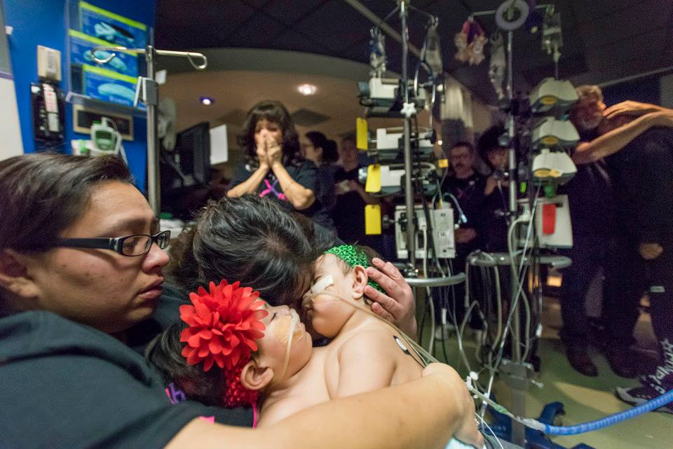
According to the University of Maryland, the survival rates of conjoined twins are often low, between 5 and 25%, and the surgery to separate them can yield difficult challenges, particularly in the case of those that share vital organs, with one twin surviving 75% of the time. At Texas Children’s Hospital, the twin daughters of Elysse Mata, Knatalye Hope and Adeline Faith, were born conjoined at the chest and abdomen, sharing, according to 3D printing and software provider Materialise, “their chest wall, lungs, pericardial sac, diaphragm, liver, intestines, colon and pelvis.” So, the surgery to separate them would, indeed, be a complex one.
With almost a year of planning, Texas Children’s Hospital surgeons, embarked upon the intricate surgery to separate the 10-month old sisters and required an accurate method for visualizing the surgery ahead of time. Nicholas Dodd, advanced visualization expert at Texas Children’s Hospital, and Dr. Jayanthi Parthasarathy of MedCAD in Dallas, collaborated to translate the infants’ CT scans, captured with a high level of contrast, into a color-coded, 3D model, using Materialise’s Mimics Innovation Suite. The surgeons were then able to 3D print a detailed model of the twins’ vital organs, including the heart, lungs, stomachs, and kidneys, and further outlining exactly how they were conjoined.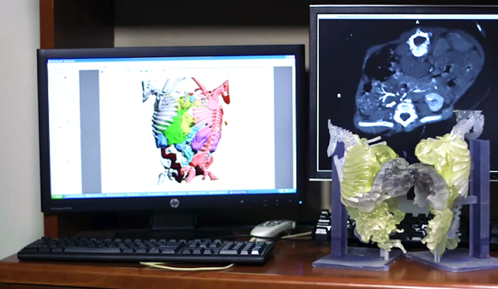
Before the surgery, the team first implanted tissue expanders into the twins’ torsos, to stretch their skin in preparation for the surgery. Then, a team of 26 doctors from 13 different specialties spent almost 30 hours to separate Knatalye Hope and Adeline Faith, doing so successfully.
Dr. Rajesh Krishnamurthy, chief of radiology research and cardiac imaging at the hospital, explains how exactly the 3D printed model was able to help cut the surgery time. He says, “Having a 3D-printed model gives you an insight into what you’re going to encounter. This type of surgical planning becomes very important when you decide to assign an organ to one twin or the other.” The quote is taken from the video below, which provides an even more in depth look into the story of the Mata twins. Let it inspire a heartfelt weekend in all who watch it!
Source: 3dprintingindustry.com/






Leave a Reply
Want to join the discussion?Feel free to contribute!